
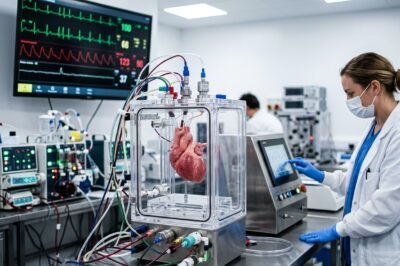
Cardiovascular disease remains the leading cause of mortality worldwide, claiming millions of lives annually despite the rapid advancement of pharmaceutical and surgical interventions. For decades, clinicians have relied on standardized protocols and anatomical generalizations to treat a pump that is as unique as a fingerprint in every patient. However, the dawn of ex vivo cardiac simulators—complex bioengineering systems that maintain a heart’s physiological function outside the human body—is set to redefine the boundaries of what is possible in precision medicine. As highlighted in recent reports by the European Medical Journal (EMJ), these simulators provide a high-fidelity environment that bridges the gap between static imaging and the dynamic, high-stakes reality of the operating room. By allowing surgeons to rehearse complex procedures on a functioning, beating heart that mimics the specific pathology of an individual patient, the medical community is moving away from reactive treatment toward a future of predictive, personalized intervention. This paradigm shift represents one of the most significant leaps in cardiology in the twenty-first century, promising to reduce surgical complications, optimize device placement, and ultimately save countless lives by mastering the complexities of the human heart before the first incision is ever made.
The Scientific Foundation of Ex Vivo Cardiac Simulators
Ex vivo cardiac simulators are not merely mechanical pumps; they are sophisticated bioreactors designed to replicate the incredibly nuanced environment of the human thoracic cavity. At their core, these systems utilize a combination of biological tissue—often porcine or bovine hearts, and increasingly, patient-specific 3D-printed synthetic analogs—integrated into a circuit that provides pulsatile flow, temperature regulation, and nutrient-rich perfusion. The goal is to maintain the heart in a state where it exhibits the same hemodynamic properties it would inside the patient. This includes replicating systolic and diastolic pressures, stroke volume, and the complex interplay of the heart valves during the cardiac cycle.
The engineering behind these devices involves high-precision sensors that monitor fluid dynamics in real-time. Researchers use blood-analog fluids that match the viscosity and density of human blood to ensure that the shear stresses on the heart walls and valves are accurately represented. By manipulating the resistance and compliance within the simulator’s tubing, scientists can recreate specific disease states, such as hypertension or aortic stenosis. This level of control allows for an unprecedented study of the heart’s mechanical behavior, providing a platform for testing how different interventions will affect the organ’s overall efficiency and structural integrity.
Advancing Precision Medicine Through Patient-Specific Modeling
One of the most compelling aspects of the EMJ report is the focus on personalization. Traditionally, a surgeon views a 2D or 3D scan of a patient’s heart and mentally projects the surgical steps required. Ex vivo simulators take this a step further by integrating patient-specific data from MRI and CT scans to create a physical or biological replica of that exact patient’s heart. This means a surgeon can encounter the specific calcification patterns, arterial twists, and valve leakages of a particular individual long before the patient is anesthetized. This approach effectively turns the simulation into a personalized dress rehearsal, reducing the likelihood of intraoperative surprises that can lead to adverse outcomes.
Furthermore, the integration of computational fluid dynamics (CFD) with ex vivo modeling allows clinicians to visualize how blood flow will change following a procedure. For instance, in complex congenital heart surgeries where the anatomy is significantly distorted, being able to simulate the flow patterns through a reconstructed vessel can prevent post-operative complications like turbulence or stagnant flow, which often lead to thrombosis. By fine-tuning the surgical plan on the simulator, the medical team can ensure that the final result provides the best possible long-term hemodynamic outcome for the patient.
Improving Surgical Outcomes and Mitigating Intraoperative Risks
The operating room is an environment where every second counts and the margin for error is razor-thin. Ex vivo simulators serve as a powerful tool for risk mitigation. In highly complex procedures, such as transcatheter aortic valve replacement (TAVR) or mitral valve repairs, the placement of a device must be perfect to avoid obstructing coronary flow or causing paravalvular leaks. By practicing these placements on a simulator, surgeons can identify the optimal angle and position for the device, customized to the patient’s unique anatomy. This pre-operative planning has been shown to significantly reduce the time a patient spends on a heart-lung bypass machine, which is a major factor in post-operative recovery and the risk of stroke.
Moreover, these simulators provide a platform for “failure testing.” Surgeons can intentionally push a procedure to its limits on the model to see where it might fail, allowing them to develop contingency plans. If a certain suture technique leads to a tear in the fragile tissue of a simulator heart, the surgeon learns to avoid that specific approach in the real surgery. This ability to make mistakes and learn from them in a consequence-free environment is a luxury that has never before been available in such high fidelity in the field of cardiac surgery, fundamentally shifting the safety profile of these operations.
The Role of Ex Vivo Systems in Medical Device Innovation
Beyond individual patient care, ex vivo cardiac simulators are revolutionizing the way medical devices are developed and approved. Historically, new heart valves, stents, and pacemakers had to undergo extensive animal testing before moving to human trials. While animal models are useful, they do not always perfectly replicate human physiology. Ex vivo simulators provide a more controlled and relevant environment for testing the durability and functionality of these devices. Engineers can run a simulator for thousands of cycles, representing years of wear and tear on a prosthetic valve, all within a few weeks in a laboratory setting.
This accelerated testing cycle not only brings life-saving technologies to market faster but also ensures they are safer. Device manufacturers can use the data from these simulations to refine their designs, making them more compatible with the wide range of anatomical variations found in the human population. The EMJ report notes that this could lead to a new generation of “smart” implants that are designed to be deployed more easily and last longer, specifically because they were optimized in a high-fidelity ex vivo environment that accounts for the dynamic mechanical stresses of a beating heart.
Transforming Medical Education and Surgical Residency
The education of the next generation of cardiac surgeons is another area poised for a massive transformation. Historically, surgical training followed the “see one, do one, teach one” model, which often involved high-pressure learning during actual human surgeries. Ex vivo simulators offer a superior alternative, allowing residents to perform full procedures on realistic tissue with the same tactile feedback they would experience in a real chest cavity. This wet-lab training is far more effective than computer-based simulations or plastic models because it involves real biological reactions and the physical complexities of handling living or preserved tissue.
Standardized testing for surgical competency could also be revolutionized. Instead of relying on subjective evaluations, residency programs can use simulator data—such as the precision of a suture, the time taken for a graft, and the resulting flow dynamics—to objectively measure a trainee’s skill level. This ensures that only those who have demonstrated mastery on the simulator move on to performing surgery on patients. As these systems become more affordable and portable, they could become a staple in every teaching hospital, democratizing high-level surgical training and raising the standard of care across the globe.
Future Directions: AI Integration and the Path to Clinical Ubiquity
Looking forward, the integration of Artificial Intelligence (AI) with ex vivo cardiac simulators promises to take personalized care to even greater heights. AI algorithms can analyze the vast amounts of data generated by the simulator—monitoring pressure changes, flow rates, and tissue stresses—to provide real-time suggestions to the surgeon. In the future, we may see a closed-loop system where the simulator, informed by AI, automatically identifies the most efficient surgical path and predicts the 10-year prognosis of the patient based on the results of the simulated procedure. This would represent the ultimate realization of precision cardiology.
However, for this technology to become a standard part of clinical practice, several challenges must be addressed. The cost of maintaining and operating high-fidelity simulators is currently high, and the ethical considerations surrounding the use of biological tissue must be managed. Furthermore, the medical community must establish clear guidelines for how simulator data should be used in clinical decision-making. Despite these hurdles, the evidence presented by the EMJ suggests that the benefits far outweigh the costs. As technology advances and costs decrease, ex vivo cardiac simulators will likely transition from specialized research tools to essential components of the surgical workflow, heralding a new era of safety, precision, and personalization in cardiovascular medicine.







































Leave a Reply