
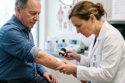
In the complex landscape of clinical dermatology, the appearance of small, bright red papules known as cherry angiomas has long been dismissed as a benign consequence of natural aging. These vascular proliferations, often referred to as Campbell de Morgan spots, are common in the general population, typically appearing after the age of 30 and increasing in frequency as the decades pass. However, a groundbreaking study recently published in the European Medical Journal (EMJ) is fundamentally reshaping our understanding of these seemingly innocuous spots. This new research provides compelling evidence that a higher-than-average prevalence of cherry angiomas may serve as a significant clinical marker for Neurofibromatosis Type 1 (NF1), a complex genetic disorder. For years, clinicians have relied on a specific set of diagnostic criteria, including cafe-au-lait spots and Lisch nodules, to identify NF1. The revelation that cherry angiomas—specifically their early onset and unusual distribution—could be linked to the NF1 mutation offers a new diagnostic window. This discovery is not merely a curiosity for medical researchers; it has profound implications for early detection, patient monitoring, and our fundamental understanding of how vascular anomalies intersect with genetic syndromes. By examining the intersection of vascular biology and genetic mutation, this EMJ report signals a shift in how we approach both common skin conditions and rare genetic diseases. ## Understanding the Clinical Significance of Cherry Angiomas in NF1. Cherry angiomas are the most common type of acquired cutaneous vascular proliferation. Histologically, they consist of dilated capillaries lined by flattened endothelial cells within the papillary dermis. While their etiology in the general population is often linked to localized increases in angiogenic factors or environmental triggers, their manifestation in patients with Neurofibromatosis Type 1 appears to follow a distinct pathological trajectory. NF1 is caused by a mutation in the NF1 gene, which encodes the protein neurofibromin. Neurofibromin acts as a negative regulator of the Ras signaling pathway. When this protein is deficient or dysfunctional, the resulting overactivity of the Ras pathway leads to uncontrolled cell growth and proliferation. The EMJ study suggests that this same pathway hyperactivity may be responsible for the premature and prolific development of cherry angiomas in NF1 patients. Unlike the general population, where these spots appear later in life, NF1 patients may exhibit multiple angiomas as early as their late teens or early twenties. This temporal shift is a critical diagnostic clue. Furthermore, the density of these lesions in NF1 cohorts far exceeds that seen in age-matched controls, suggesting that the genetic environment created by the NF1 mutation is uniquely fertile for vascular tumor growth. ## Breaking Down the Research: Methodology and Key Findings from EMJ. The study featured in the EMJ involved a comprehensive cross-sectional analysis of patients diagnosed with NF1 compared to a control group of healthy individuals. Researchers meticulously documented the number, size, and anatomical distribution of cherry angiomas across various age groups. One of the most striking findings was that cherry angiomas were present in nearly 50 percent of NF1 patients under the age of 40, a demographic where such lesions are typically rare. The methodology also included dermatoscopic evaluations, which revealed that angiomas in NF1 patients often displayed a more intense vascular pattern than those found in the general public. Additionally, the researchers utilized molecular testing to confirm that the endothelial cells within these specific angiomas exhibited the same ‘second hit’ mutations often seen in neurofibromas. This finding provides a direct molecular bridge between the genetic disorder and the vascular symptom. The study also noted that the presence of cherry angiomas was frequently correlated with other vascular abnormalities, such as glomus tumors, which are already known to be associated with NF1. This suggests that cherry angiomas are part of a broader spectrum of vascular manifestations within the NF1 phenotype rather than an isolated finding. ## The Genetic Blueprint: How NF1 Mutations Influence Vascular Lesions. To understand why cherry angiomas are linked to NF1, one must look deep into the molecular machinery of the cell. The NF1 gene is a large and complex gene located on chromosome 17. Its primary product, neurofibromin, is a GTPase-activating protein (GAP) that facilitates the conversion of active Ras-GTP to inactive Ras-GDP. When neurofibromin is absent, Ras remains in its active state, triggering downstream cascades including the MAPK and PI3K/mTOR pathways. These pathways are central to angiogenesis—the process of new blood vessel formation. Specifically, the activation of the mTOR pathway has been identified as a key driver in the proliferation of endothelial cells that form cherry angiomas. In the context of NF1, every cell in the body carries one mutated copy of the gene. When a somatic mutation occurs in the second copy within a vascular endothelial cell, the inhibitory control on Ras is lost, leading to the rapid formation of a cherry angioma. This ‘two-hit’ hypothesis, originally proposed for neurofibromas, appears to apply equally to these vascular lesions. This genetic insight is revolutionary because it suggests that cherry angiomas are not just associated with NF1 but are a direct manifestation of the genetic defect itself, functioning as a cutaneous signpost of internal molecular signaling gone awry. ## Statistical Insights: Prevalence Rates and Diagnostic Accuracy. Statistics from the EMJ report provide a stark contrast between NF1-affected individuals and the general population. In a cohort of 200 NF1 patients, the mean number of cherry angiomas per patient was significantly higher (averaging 12-15 lesions) compared to the control group (averaging 2-4 lesions in similar age brackets). More importantly, the sensitivity of using cherry angiomas as a secondary diagnostic criterion was found to be notably high in adult populations. While they do not replace the classic NIH criteria—such as six or more cafe-au-lait macules or two or more neurofibromas—they provide an additional layer of diagnostic confidence. In cases where a patient may only partially meet existing criteria, the presence of multiple, early-onset cherry angiomas could tip the balance toward a definitive NF1 diagnosis. The study calculated that the presence of more than 10 cherry angiomas in a patient under the age of 30 has a high predictive value for an underlying Ras-pathway disorder. This statistical evidence is vital for general practitioners and dermatologists who are often the first to see these patients. By recognizing these ‘red flags,’ clinicians can initiate genetic testing earlier, leading to better management of the more serious complications associated with NF1, such as optic gliomas or malignant peripheral nerve sheath tumors. ## Comparative Analysis: Differentiating Sporadic Cherry Angiomas from NF1-Associated Lesions. For a clinician, the challenge lies in distinguishing between a ‘normal’ cherry angioma and one associated with NF1. While they look identical to the naked eye, the EMJ study highlights several differentiating factors. First is the age of onset; sporadic cherry angiomas are hallmarks of cutaneous aging, whereas NF1-associated lesions appear much earlier. Second is the distribution. While sporadic angiomas are typically scattered on the trunk, NF1-associated angiomas often appear in higher clusters and may even appear on the extremities more frequently than expected. Third is the association with other NF1 markers. If a patient presents with multiple cherry angiomas and a history of learning disabilities, scoliosis, or even a single plexiform neurofibroma, the suspicion of NF1 should be significantly heightened. The EMJ researchers also suggested that the rate of appearance is a factor; NF1 patients often report the sudden ‘eruptive’ appearance of dozens of new angiomas over a short period, a phenomenon rarely seen in the general population unless triggered by specific medications or chemical exposures. Understanding these nuances allows for a more sophisticated clinical assessment that moves beyond simple observation to proactive genetic screening. ## Future Directions in Personalized Medicine and Early Diagnosis. The link between cherry angiomas and NF1 opens several new avenues for medical advancement. Primarily, it underscores the importance of the skin as a window into systemic health. In the era of personalized medicine, identifying such accessible biomarkers is crucial. Future research may focus on whether the number of cherry angiomas correlates with the severity of the NF1 phenotype or the risk of developing specific internal tumors. If a high burden of angiomas signals a more aggressive form of the disease, they could serve as a non-invasive prognostic tool. Furthermore, this discovery may lead to new therapeutic interventions. Since the mTOR pathway is implicated in both neurofibromas and cherry angiomas, the use of topical mTOR inhibitors could potentially treat both types of lesions simultaneously. As we continue to refine the diagnostic criteria for NF1, the inclusion of vascular lesions like cherry angiomas is likely to be discussed at the international level. This study serves as a reminder that even the most common and benign-looking skin features can hold the key to unlocking the secrets of complex genetic syndromes. For the thousands of individuals living with NF1, these insights offer hope for faster diagnosis and a more comprehensive approach to their lifelong care. The European Medical Journal has highlighted a vital connection that bridges the gap between everyday dermatology and high-level genetics, proving that in medicine, it pays to look closer at the small things.
Redefining Dermatological Markers: The Surprising Link Between Cherry Angiomas and Neurofibromatosis Type 1
Posted by
Stories




-

Mithila Palkar: The Relatable Queen of Indian Web Series and Her Meteoric Rise
-

Wamiqa Gabbi: The Meteoric Rise of Indian Web Series’ Most Versatile Leading Lady
-

Triptii Dimri Biography: The Evolution of India’s Newest Cinematic Sensation and Web Series Star
-

Wamiqa Gabbi: The Rise of India’s New Digital Icon and the Future of OTT Stardom
-

Mithila Palkar: The Unstoppable Evolution of India’s Digital Darling and Her Journey to the Silver Screen
-

Mithila Palkar: The Comprehensive Journey of India’s Web Series Sensation and Digital Pioneer
-

Mithila Palkar: The Relatable Queen of Indian Web Series and Her Inspiring Journey to Stardom
-

Sobhita Dhulipala Biography: The Meteoric Rise of India’s Most Versatile Web Series Star
-

Mithila Palkar: The Unstoppable Journey of India’s Digital Darling and Web Series Queen
-

Triptii Dimri: The Complete Biography of India’s Newest Cinematic Sensation and Web Series Icon
-

Wamiqa Gabbi: The Ethereal Rise of India’s Most Versatile Web Series Star
-

Sobhita Dhulipala: The Unstoppable Rise of India’s Most Versatile Web Series Icon
-

Wamiqa Gabbi: The Meteoric Rise of the New Queen of Indian Web Series and Her Unstoppable Journey to Stardom
-

Triptii Dimri: The Meteoric Rise of India’s New Cinema Sweetheart and Web Series Icon
-

Wamiqa Gabbi: The Versatile Queen of Indian OTT and Her Phenomenal Rise to Stardom
-

Tripti Dimri Biography: The Rise of India’s New National Crush and Her Journey from Laila Majnu to OTT Stardom
-

Wamiqa Gabbi Biography: The Meteoric Rise of India’s Newest OTT Sensation and Versatile Powerhouse
-

Wamiqa Gabbi Biography: The Meteoric Rise of the Digital Queen of Indian Web Series
-

The Rise of Wamiqa Gabbi: A Deep Dive into the Journey of India’s Most Versatile OTT Sensation
-

The Meteoric Rise of Triptii Dimri: Biography, Acting Prowess, and the Journey of India’s Newest Cinema Sensation
-

Wamiqa Gabbi Biography: The Meteoric Rise of the Digital Queen and Her Unmatched Acting Prowess

Launching Soon: The Future of News with Our E-Newspaper
In the ever-evolving landscape of media and technology, we are thrilled to announce the upcoming launch of our innovative e-newspaper, set to redefine the way news is consumed in the digital age. Embracing the convenience and accessibility that the digital world offers, our e-newspaper aims to deliver real-time news updates, insightful articles, and interactive features directly to your devices. With a commitment to journalistic integrity and a passion for storytelling, we are dedicated to keeping you informed, engaged, and connected, no matter where you are. Stay tuned for the launch of our e-newspaper, where the future of news awaits at your fingertips.
Recent News
- Breakthrough in Pain Management: How Cancer Medications are Repurposing for Chronic Nerve Pain Relief

- Revolutionizing Carbon Capture: How IIT Gandhinagar is Leveraging AI to Turn CO₂ into Clean Energy

- India’s Economic Resurgence: A Comprehensive Analysis of the 7.8 Percent Q1 Growth Beat

- Redefining Dermatological Markers: The Surprising Link Between Cherry Angiomas and Neurofibromatosis Type 1
- 150 Years of the Grand Canyon: From John Wesley Powell’s 1869 Discovery to Modern Geological Revaluations

- The Oncologist’s Manifesto: A Comprehensive Breakdown of Prostate Cancer Awareness and Modern Treatment

Politics
- India’s Economic Resurgence: A Comprehensive Analysis of the 7.8 Percent Q1 Growth Beat
- Beyond the Ballot: An In-Depth Analysis of the RSS-BJP Power Dynamic and Mohan Bhagwat’s Evolving Rhetoric

- India and Uzbekistan Elevate Relations to Comprehensive Strategic Partnership: A New Era of Central Asian Diplomacy

- The Great Cleansing: Xi Jinping’s Purge Reaches the Commanders of Doklam and Galwan

- Fed Chair Kevin Warsh at Jackson Hole: ‘We have work to do’ on inflation – A Turning Point for Markets

- India’s Strategic Shift: Decoding Ex-CDS Chauhan’s Insights on Skardu vs Sargodha Airbases

Categories
- Art & Culture 100
- Auto 85
- Beauty 15
- Blog 251
- Books 7
- Business 359
- Celebrities 217
- Crime Files 139
- Democracy 308
- Education 84
- Entertainment 154
- Environment 133
- epaper 1
- Fashion 37
- Food 58
- Gadgets 86
- Guide 150
- Handicrafts 10
- Health 263
- Healthy Living 107
- History 5
- incidents 156
- Lifestyle 31
- Music 5
- Photography 15
- Politics 425
- Science & Technology 160
- Sports 228
- Tech 123
- Television 2
- Travel 68
- Travel & Tourism 30
- Trends 40
- Weather 6
- Web Series 96
- Workout 4
- World 651
Leave a Reply