
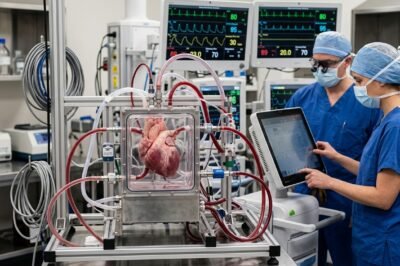
The cardiovascular landscape is on the precipice of a radical transformation, moving away from a one-size-fits-all approach toward a highly refined era of precision medicine. For decades, the intricacies of the human heart have presented a unique challenge to clinicians, where the margin for error is razor-thin and the physiological variations between individuals are vast. According to recent insights from the European Medical Journal (EMJ), the emergence of ex vivo cardiac simulators is set to bridge the critical gap between theoretical surgical planning and actual clinical outcomes. These sophisticated systems allow for the reanimation and study of cardiac tissues outside the human body, providing a high-fidelity environment that mimics the complex hemodynamics of a living patient. This breakthrough is not merely an incremental improvement; it represents a paradigm shift in how surgeons prepare for complex procedures and how medical devices are validated. By utilizing patient-specific data to drive these simulations, the medical community can now visualize and test interventions with a level of accuracy that was previously confined to the realm of science fiction. The implications for patient safety, surgical success rates, and the acceleration of medical innovation are profound, marking a new chapter in the fight against global cardiovascular disease.
The Evolution of Cardiac Simulation Technology
To understand the significance of ex vivo simulators, one must first look at the history of cardiac modeling. Historically, surgeons relied on static imaging like X-rays, and later, 2D and 3D echocardiography or CT scans to map out surgeries. While these tools provided vital structural information, they lacked the dynamic, functional dimension of a beating heart. The introduction of computational fluid dynamics (CFD) allowed for digital modeling of blood flow, but these mathematical simulations often struggle to account for the organic variability and mechanical properties of living tissue. The ex vivo approach changes this by integrating physical heart components—either biological tissue or advanced bio-synthetic materials—into a mechanical circuit that replicates the human circulatory system.
The EMJ report highlights that these simulators have evolved from simple pump systems to multi-chambered apparatuses capable of simulating specific pathologies, such as mitral valve regurgitation or aortic stenosis. By fine-tuning the pressure, flow rate, and viscosity of the circulating fluid, researchers can recreate the exact conditions of a patient’s cardiovascular system. This level of detail is crucial because it allows for the observation of how heart tissue reacts to physical stress in real-time. The ability to monitor myocardial contractility and valvular function under controlled laboratory conditions provides a wealth of data that simply cannot be obtained during a live operation where the primary focus is on the patient’s immediate survival.
Mechanisms of Ex Vivo Heart Simulators
The technical architecture of an ex vivo cardiac simulator is a feat of modern engineering. At its core, the system consists of a pulsatile pump that acts as the ventricle, a series of reservoirs representing the atria, and a complex network of tubing that mimics the resistance and compliance of the systemic and pulmonary vasculature. One of the most critical components is the use of a blood-mimicking fluid, which must possess the same Newtonian or non-Newtonian properties as human blood to ensure that the shear stress on valve leaflets and vessel walls is accurately represented. The integration of high-speed cameras and pressure sensors allows for the collection of granular data at every stage of the cardiac cycle.
Furthermore, the use of porcine or bovine hearts in these simulators has become a standard practice for testing surgical techniques. These biological hearts are decellularized or specially treated to maintain their mechanical integrity while allowing surgeons to practice incisions, suturing, and the placement of prosthetic devices. The EMJ analysis points out that these biological models are increasingly being augmented with 3D-printed components derived from actual patient MRI scans. This hybrid approach ensures that the simulator is not just a general representation of a heart, but a bespoke model of a specific patient’s unique anatomy, including any congenital defects or calcifications that might complicate a procedure.
Bridging the Gap in Personalized Medicine
Personalized medicine is the ultimate goal of modern healthcare, and in cardiology, this means tailoring an intervention to the specific structural and functional nuances of a patient’s heart. Ex vivo simulators are the key to unlocking this potential. For instance, in the case of Transcatheter Aortic Valve Replacement (TAVR), the fit of the prosthetic valve is paramount. A slightly mismatched valve can lead to paravalvular leaks or even catastrophic displacement. By using a patient-specific ex vivo simulator, clinicians can test different valve sizes and types before the patient even enters the operating room, identifying the optimal configuration that ensures a perfect seal and long-term durability.
This “pre-flight” testing phase is revolutionary. It allows the surgical team to anticipate complications and refine their strategy in a zero-risk environment. The EMJ notes that this is particularly beneficial for complex cases where conventional guidelines may not apply. For patients with multiple comorbidities or atypical anatomy, the simulator serves as a diagnostic tool that goes beyond imaging, offering a physical trial run. This reduces the time the patient spends under anesthesia and minimizes the likelihood of mid-procedure adjustments, which are often the source of surgical complications and increased recovery times.
Impact on Surgical Training and Innovation
Beyond individual patient care, ex vivo simulators are transforming the educational landscape for the next generation of cardiothoracic surgeons. The traditional “see one, do one, teach one” model is being replaced by a more rigorous, simulation-based curriculum. Residents can now perform high-stakes procedures hundreds of times on a simulator before ever touching a human heart. This deliberate practice allows for the mastery of fine motor skills and the development of clinical intuition in a way that was previously impossible. The ability to simulate rare emergencies—such as a sudden ventricular rupture or acute valve failure—prepares surgeons to respond with calm precision when these events occur in the real world.
In the realm of medical device development, these simulators are accelerating the path from concept to clinic. Manufacturers of stents, artificial valves, and ventricular assist devices (VADs) can use ex vivo systems to conduct rigorous stress tests and durability studies. This high-fidelity testing can often provide enough evidence to streamline the regulatory approval process, as it offers a more realistic assessment of device performance than traditional bench testing. By reducing the reliance on animal models, which often have different anatomical proportions than humans, ex vivo simulators provide a more direct and ethically sound route to innovation.
Economic and Clinical Outcomes
The implementation of ex vivo simulation technology also carries significant economic implications. While the initial setup cost for these systems can be high, the long-term savings are substantial. By reducing the rate of re-operations and post-surgical complications, hospitals can significantly lower the cost of care for cardiovascular patients. Complications such as strokes or heart failure following a botched valve replacement are not only devastating for the patient but are also incredibly expensive for the healthcare system to manage. The EMJ highlights that data-driven surgical planning leads to shorter hospital stays and faster returns to productive life for patients.
From a clinical perspective, the statistics are equally compelling. Early studies suggest that the use of patient-specific models in surgical planning can improve procedural success rates by up to 15% in complex cases. Furthermore, the ability to optimize drug delivery systems using these simulators—testing how localized treatments circulate through the heart’s unique flow patterns—is opening new doors for non-surgical interventions. As healthcare systems move toward value-based care models, the integration of simulation technology will likely become a benchmark for quality and safety in cardiovascular departments worldwide.
Future Implications and the Horizon of Cardiac Care
Looking forward, the future of ex vivo cardiac simulators lies in their integration with artificial intelligence and machine learning. By feeding the massive amounts of data generated during simulations into AI algorithms, we can begin to predict long-term outcomes with unprecedented accuracy. We may soon reach a point where an AI can analyze a patient’s simulator performance and suggest the surgical approach with the highest statistical probability of success. Additionally, as 3D bioprinting technology matures, we may see the transition from using porcine hearts to using fully synthetic, bio-printed human hearts that contain the patient’s own cells, eliminating the need for biological tissue donors altogether.
The EMJ’s report on ex vivo cardiac simulators serves as a clarion call for the medical community to embrace these technological advancements. We are moving toward a future where “zero-risk” surgery is the standard, and where every cardiovascular intervention is as unique as the patient receiving it. As these systems become more accessible and integrated into standard clinical workflows, the mystery of the human heart will continue to be unraveled, leading to longer, healthier lives for millions of people. The era of personalized cardiovascular care is no longer a distant dream; it is being built, beat by beat, in the simulation labs of today.






































Leave a Reply