
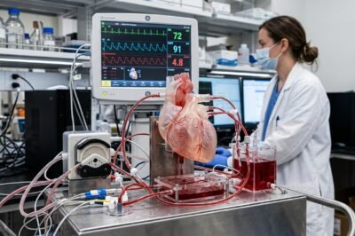
The human heart, a tireless pump beating approximately 100,000 times a day, has long remained one of the most challenging organs to treat with absolute precision due to its complex fluid dynamics and varied anatomical structures. Despite decades of advancements in pharmacology and surgical techniques, cardiovascular disease remains the leading cause of mortality globally, necessitating a more refined approach to intervention. However, a paradigm shift is currently occurring, driven by the emergence of ex vivo cardiac simulators—sophisticated mechanical and biological systems that replicate the heart’s function outside the living body. Recent discussions and studies highlighted by the European Medical Journal (EMJ) emphasize how these technologies are not merely academic curiosities but are becoming pivotal tools in personalizing cardiovascular care. By allowing clinicians to test interventions on a patient’s unique physiological profile without the inherent risks of live surgery, these simulators are bridging the gap between theoretical medicine and tactile surgical excellence. This transition marks the end of the “trial and error” era in cardiology, ushering in a future where every incision, stent placement, and valve replacement is pre-validated in a high-fidelity synthetic environment. The potential for these devices to reduce procedural complications and improve long-term patient outcomes is immense, representing a true convergence of mechanical engineering, computer science, and biological medicine.
Understanding Ex Vivo Cardiac Simulators: The Mechanics of Mock Circulation
Ex vivo cardiac simulators, often referred to in the engineering community as mock circulatory loops (MCLs), are complex systems designed to mimic the hemodynamic conditions of the human circulatory system with extreme accuracy. At their core, these systems consist of a series of reservoirs, pulsatile pumps, and adjustable valves that replicate the heart’s four chambers and the systemic and pulmonary vascular beds. The primary objective of these simulators is to maintain realistic pressure-flow relationships, which are governed by critical physical parameters such as compliance—the ability of the vessels to expand and contract—and peripheral resistance—the friction encountered by blood as it flows through the smaller vessels. By adjusting these physical variables, researchers can simulate a vast range of physiological states, from the resting heart rate of a healthy athlete to the high-pressure environment of a patient with chronic hypertension or the weakened, inefficient flow characteristic of congestive heart failure.
The sophistication of these simulators has advanced significantly with the integration of microprocessor-controlled pulsatile pumps that can replicate the exact rhythmic contraction of the cardiac muscle. Unlike older steady-flow pumps, modern pulsatile systems produce the systolic and diastolic phases essential for testing dynamic medical devices like artificial heart valves or ventricular assist devices (VADs). The fluid used in these systems is often a carefully formulated blood-analog, such as a glycerin-water mixture, which is calibrated to match the specific viscosity and density of human blood at body temperature. This attention to detail ensures that the shear stresses and turbulence patterns observed within the simulator are representative of what would occur within a patient’s own vasculature. According to reports in the EMJ, this high-fidelity replication provides engineers and clinicians with invaluable data on flow dynamics and wall stress that was previously unobtainable without invasive human trials or potentially misleading animal models.
Personalized Medicine: From High-Resolution Imaging to Physical Heart Replicas
The most transformative aspect of ex vivo cardiac simulators is their ability to incorporate patient-specific data, turning the concept of personalized medicine into a physical reality. This meticulous process begins with high-resolution medical imaging, where computed tomography (CT) or magnetic resonance imaging (MRI) scans are used to create a detailed three-dimensional digital reconstruction of a patient’s unique anatomy. This “digital twin” captures the nuances of the patient’s cardiac structure, including the specific shape of the chambers, the thickness of the myocardial walls, and the orientation of the major vessels. This data is then converted into a physical model using advanced additive manufacturing techniques, such as 3D printing or multi-material stereolithography. By using flexible, biocompatible resins that mimic the Young’s modulus and tactile properties of human cardiac tissue, engineers can create a physical replica of a specific patient’s heart.
This level of personalization allows for a “bench-to-bedside” planning phase that is unprecedented in the history of surgery. For instance, in complex cases of Transcatheter Aortic Valve Implantation (TAVI), a simulator can be used to test several different valve sizes and deployment positions to see which one provides the optimal hemodynamic fit while minimizing the risk of paravalvular leakage or conduction disturbances. This pre-operative testing is particularly crucial for patients with atypical anatomy or congenital heart defects who might otherwise be considered high-risk for standard procedures. By identifying potential complications on the physical simulator before the patient ever enters the operating room, surgical teams can refine their strategy, reduce the time the patient spends under anesthesia, and significantly lower the probability of intraoperative emergencies. The EMJ analysis suggests that as 3D printing costs continue to decline, this patient-specific simulation could become the standard protocol for all complex cardiovascular interventions.
The Role of Simulators in Accelerating Medical Device R&D and Safety
Beyond individual patient care, ex vivo cardiac simulators are revolutionizing the entire research and development cycle for the medical device industry. Historically, the path from a new stent or valve design to clinical approval has been extraordinarily long and fraught with difficulty, often relying heavily on animal models that, while useful, do not perfectly replicate human-specific anatomy or the unique fluid dynamics of the human heart. Simulators provide a highly repeatable and controllable environment where new devices can be tested under extreme conditions—such as severe hypertension or rapid tachycardia—that would be unethical or impossible to study in living subjects. This enables manufacturers to identify potential failure points or structural weaknesses early in the design process, leading to the creation of safer and more durable products.
Furthermore, the EMJ highlights that these simulators are instrumental in the development of the next generation of “smart” implants. As the medical community moves toward an era of bio-electronic devices that can monitor and respond to changes in a patient’s condition, the ability to test these sensors in a high-fidelity hemodynamic environment is essential. Simulators can be used to calibrate pressure sensors, test the durability of lead wires, and ensure that the software algorithms governing the device can accurately interpret physiological signals in real-time. This synergy between mechanical simulation and digital health is paving the way for autonomous cardiac supports and advanced pacing technologies that can adapt their output based on the simulated needs of the patient. By providing a rigorous testing ground, simulators ensure that these complex technologies are proven safe and effective before they ever reach the clinical trial phase.
Educational Paradigms: The “Flight Simulator” for Cardiac Surgeons
The pedagogical value of ex vivo cardiac simulators for the next generation of medical professionals cannot be overstated. Traditional surgical training has long followed the “see one, do one, teach one” model, which, while effective, inherently places a burden on patient safety during the early stages of a surgeon’s learning curve. Ex vivo simulators offer a sophisticated “flight simulator” for cardiac surgery, providing a risk-free environment where residents and fellows can practice complex procedures repeatedly until they achieve technical mastery. These systems can be programmed to simulate specific surgical complications, such as a ruptured coronary vessel or a sudden drop in systemic pressure, forcing the trainee to react under pressure and develop the critical thinking skills and muscle memory necessary for success in the operating room.
Moreover, the integration of sensors within the simulator allows for the objective assessment of surgical skill. Advanced platforms can track the amount of force applied by the surgeon during suturing, the precision of their instrument placement, and the overall efficiency of their movements. This data-driven approach to training ensures that only those who meet a high, measurable standard of technical proficiency are cleared to perform procedures on live patients. As surgical techniques continue to evolve toward minimally invasive and robotic-assisted methods, simulators will remain an indispensable tool for helping even experienced surgeons transition to new technologies without compromising the quality of care. The EMJ notes that medical schools and teaching hospitals that incorporate high-fidelity simulation into their curricula report higher levels of trainee confidence and a marked reduction in procedural errors among their graduates.
Economic and Ethical Implications: A Sustainable Future for Healthcare
While the initial investment in ex vivo cardiac simulation technology and 3D printing infrastructure can be significant, the long-term economic benefits for healthcare systems are substantial. By improving the precision of interventions and reducing the rate of post-operative complications, these systems can significantly lower the costs associated with hospital readmissions, prolonged intensive care stays, and long-term disability. A single avoided complication in a complex cardiac case can save a healthcare system tens of thousands of dollars, quickly offsetting the cost of the simulator. Furthermore, by streamlining the medical device approval process through more accurate pre-clinical testing, simulators can bring life-saving technologies to market faster, providing a faster return on investment for companies and expanding access to care for patients globally.
Ethically, the move toward ex vivo simulation addresses growing global concerns regarding the use of animal models in medical research. While animal testing has been a cornerstone of biological science for centuries, it is increasingly scrutinized for its ethical implications and its limited predictive value for certain human-specific outcomes. Ex vivo simulators provide a more accurate representation of human-specific fluid dynamics, anatomy, and tissue behavior, potentially reducing the number of animals required for cardiovascular research. This alignment of scientific accuracy and ethical responsibility is a major theme in modern bioethics, positioning simulators as a more humane and scientifically superior alternative. The transition to these models represents a commitment to the “3Rs” of animal research—replacement, reduction, and refinement—while simultaneously enhancing the quality of data available to scientists.
Future Outlook: Integrating Artificial Intelligence and Virtual Reality
Looking toward the future, the convergence of ex vivo simulators with artificial intelligence (AI) and virtual reality (VR) promises to further redefine the landscape of cardiovascular medicine. Imagine a scenario where a surgeon wears a VR headset to perform a practice procedure on a physical, haptic-enabled simulator, with AI providing real-time feedback on their performance and suggesting the optimal tool paths based on a global database of thousands of successful outcomes. This hybrid approach would combine the tactile feedback of physical simulation with the analytical power of digital intelligence, creating a training and planning environment that is far greater than the sum of its parts. As sensors become even more miniaturized and computational power continues to grow, we may see the development of “digital-physical twins” that update in real-time based on a patient’s wearable sensor data, allowing for continuous monitoring and simulation of their condition over years.
In conclusion, the insights shared by the EMJ regarding ex vivo cardiac simulators point toward a new era of precision medicine and clinical excellence. These devices are no longer just laboratory prototypes; they are becoming essential components of a modern healthcare ecosystem that prioritizes patient safety, clinical efficiency, and ethical research practices. By providing a high-fidelity, patient-specific platform for planning, testing, and training, ex vivo simulators are ensuring that the heart—the very engine of human life—is treated with the highest degree of care and technical precision possible. As the technology matures and becomes more widely adopted across global medical centers, it will undoubtedly become the standard of care, transforming the landscape of cardiology and improving the lives of millions of patients for generations to come.






































Leave a Reply