
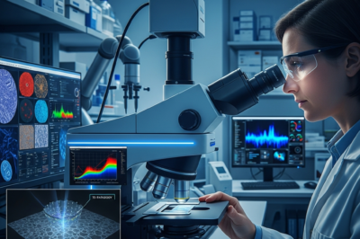
The landscape of modern medicine is currently witnessing a tectonic shift as we move from traditional diagnostic methodologies toward a sophisticated, data-driven era known as 7D pathology. At the heart of this revolution lies a breakthrough in optical engineering: metasurface polarimetry. This cutting-edge technology, recently highlighted in major scientific reports, promises to transform how clinicians view, analyze, and diagnose tissue samples. By capturing more information than ever before from a single microscopic slide, 7D pathology is not just an incremental improvement; it is a fundamental leap forward that integrates spatial, spectral, and polarization data into a multidimensional diagnostic framework. For decades, pathologists have relied on two-dimensional stained tissue sections to make life-altering decisions. However, the emergence of metasurface-based imaging systems is now allowing scientists to extract hidden signatures of disease that were previously invisible to the human eye and standard cameras. This transition represents a convergence of nanophotonics, artificial intelligence, and clinical oncology, marking a new chapter in the pursuit of precision medicine and personalized patient care.
The Evolution of Pathology: From Glass Slides to 7D Imaging
To understand the magnitude of the shift toward 7D pathology, one must first appreciate the history of the field. For over a century, the ‘gold standard’ of diagnosis has been the hematoxylin and eosin (H&E) stain, viewed under a conventional brightfield microscope. While effective, this method is inherently limited to spatial morphology. Digital pathology moved this process into the computer age, allowing for remote viewing and rudimentary image analysis. However, even digital pathology remained largely confined to three dimensions: two spatial dimensions and color (RGB) intensity. The concept of 7D pathology expands this significantly. It incorporates three spatial dimensions (X, Y, and Z-stacking), time-resolved measurements, spectral data (multiple wavelengths), and, most importantly, the full polarimetric state of light. Metasurface polarimetry acts as the technical engine for this expansion. Metasurfaces are ultra-thin, nanostructured surfaces that can manipulate light at a sub-wavelength scale. Unlike bulky traditional polarimeters that require rotating parts or multiple filters, a single metasurface can capture the entire Stokes vector of light in a single snapshot. This efficiency is what makes 7D pathology clinically viable, offering a level of detail that could previously only be achieved in high-end physics laboratories.
The Power of Metasurface Polarimetry in Tissue Analysis
Metasurfaces are often referred to as ‘flat optics.’ They consist of arrays of nanopillars or nano-antennas designed to interact with light in specific ways. In the context of pathology, when light passes through a biopsy specimen, its polarization state changes depending on the microstructural properties of the tissue. For instance, collagen fibers, which play a critical role in cancer progression and wound healing, are birefringent. Traditional microscopes ignore this polarization information, effectively discarding half of the data available in the light field. Metasurface polarimetry captures this data with unprecedented precision. By analyzing the Mueller matrix—a mathematical representation of how a sample affects polarized light—researchers can determine the density, orientation, and heterogeneity of cellular structures. This is particularly useful in identifying the ‘pre-cancerous’ changes in the extracellular matrix that occur before visible morphological changes appear. Because metasurfaces are manufactured using semiconductor fabrication techniques, they are compact and can be integrated directly into existing microscope objectives or even portable diagnostic devices, making this advanced technology accessible to hospitals worldwide.
Unlocking New Dimensions: What 7D Pathology Reveals
The ‘7D’ in 7D pathology refers to a multidimensional data cube that provides a holistic view of the biological specimen. While the exact definitions of the dimensions can vary, they generally include the three spatial axes, spectral frequency, temporal dynamics, and the complex degrees of light polarization (amplitude and phase). When these seven dimensions are combined, the resulting diagnostic image is far more than a simple photograph; it is a comprehensive map of the tissue’s physical and chemical state. For example, in breast cancer diagnostics, 7D imaging can differentiate between benign architectural distortions and invasive ductal carcinoma by mapping the specific alignment of collagen types. It can also assist in ‘stain-free’ pathology. Because polarization is sensitive to the refractive index of cellular components, clinicians can potentially observe cellular details without the need for chemical dyes, which can sometimes distort the tissue or lead to inconsistent results across different laboratories. This high-dimensional data is also a goldmine for machine learning algorithms, which can be trained to recognize ‘7D signatures’ of specific diseases, leading to faster and more objective diagnostic outputs.
Clinical Applications: Accuracy in Cancer Detection and Margin Analysis
One of the most critical applications of metasurface-enabled 7D pathology is in the field of surgical oncology. When a surgeon removes a tumor, they must ensure that the ‘margins’ are clear—meaning no cancer cells are left at the edge of the excised tissue. Currently, this involves a ‘frozen section’ analysis that can take 20 to 30 minutes while the patient remains under anesthesia. The precision of 7D polarimetry could allow for near-instantaneous margin analysis. By scanning the tissue with a metasurface-enhanced probe, the system could highlight areas of abnormal polarization that indicate residual cancerous cells. Furthermore, in the diagnosis of fibrosis, kidney disease, and neurodegenerative disorders, the ability to quantify structural changes at the nanoscale is invaluable. The technology provides a quantitative ‘biomarker’ rather than a qualitative ‘opinion,’ reducing the intra-observer variability that often plagues traditional pathology. As we move toward a future where treatment is tailored to the individual, having a 7D profile of a patient’s biopsy will allow oncologists to predict how a tumor might respond to specific therapies, such as immunotherapy or targeted chemotherapy.
Overcoming the Technological Hurdles of High-Dimensional Imaging
Despite its promise, the transition to 7D pathology is not without challenges. The primary hurdle is the sheer volume of data generated. A single 7D scan of a tissue slide can produce terabytes of information, requiring massive storage capacities and high-performance computing clusters to process. Furthermore, the integration of metasurfaces into clinical workflows requires rigorous validation. Medical devices must undergo extensive testing to ensure that the polarization data is consistent across different machines and environments. There is also the ‘black box’ problem associated with the AI used to interpret 7D data; pathologists must be able to understand why an algorithm flagged a specific area as malignant. However, the industry is already responding to these needs. New data compression techniques and ‘edge computing’—where the data is processed directly on the imaging device—are making 7D pathology more manageable. Additionally, international collaborations are working to standardize polarimetric metrics, ensuring that a ‘polarization signature’ in London is interpreted the same way in Mumbai or New York.
The Economic and Global Impact of Next-Generation Diagnostics
From an economic perspective, 7D pathology and metasurface technology represent a significant opportunity for cost savings in the long term. While the initial investment in new hardware is substantial, the reduction in diagnostic errors, the elimination of expensive chemical reagents through stain-free imaging, and the speed of automated analysis can drastically lower the cost per patient. In developing nations, where trained pathologists are in short supply, a metasurface-based diagnostic tool could serve as a powerful ‘triage’ system. A technician could capture the 7D data, and an AI model could provide an initial screening, flagging only the most complex cases for human review. This democratizes high-end healthcare, bringing the power of advanced physics to the point of care. As the manufacturing of metasurfaces becomes more commoditized through existing silicon foundries, we can expect the price of these sensors to drop, much like the trajectory of CMOS camera sensors in smartphones.
Conclusion: A New Dawn for Personalized Medicine
The integration of metasurface polarimetry into the realm of pathology marks a definitive turning point in medical science. By moving into the seventh dimension of imaging, we are no longer looking at tissues as static, flat images, but as complex, dynamic environments rich with hidden information. This technology provides the bridge between the macroscopic world of clinical symptoms and the nanoscopic world of molecular changes. As 7D pathology continues to evolve, it will undoubtedly lead to earlier detections, more accurate diagnoses, and ultimately, higher survival rates for patients facing complex diseases. The journey from a simple glass slide to a 7D digital twin of a human cell is a testament to human ingenuity and our relentless pursuit of understanding the fundamental building blocks of life. The future of diagnostics is no longer just about seeing; it is about perceiving the invisible, and metasurface polarimetry is the lens that makes it possible.


Leave a Reply